
What Can an MRI Show After a Suspected Neonatal Stroke?
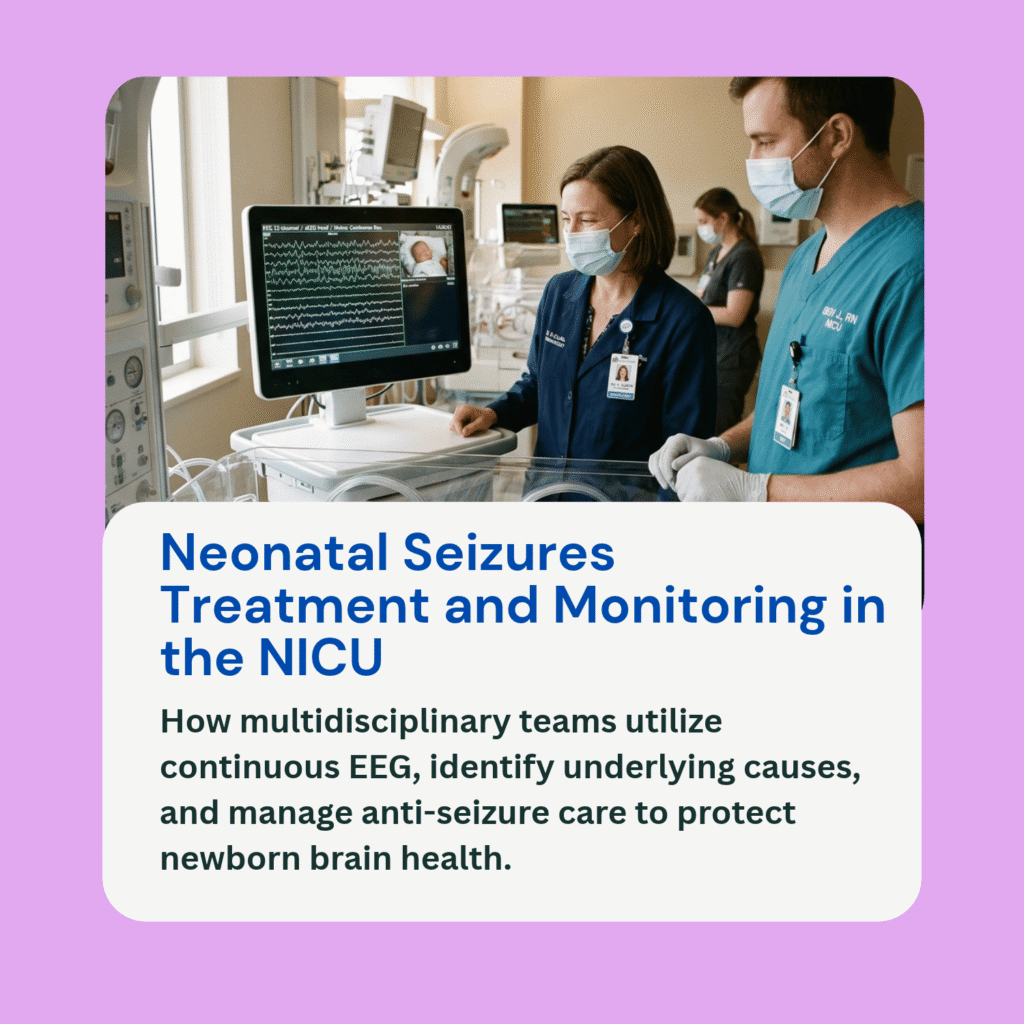
When a newborn displays early subtle focal signs, such as repetitive twitching on one side of the body or localized electrographic seizure spikes on continuous brain monitoring, neonatologists investigate for potential vascular events. While global oxygen deprivation leads to diffuse brain injury, a perinatal arterial ischemic stroke occurs when blood flow is blocked in a specific cerebral vessel, most commonly the middle cerebral artery.
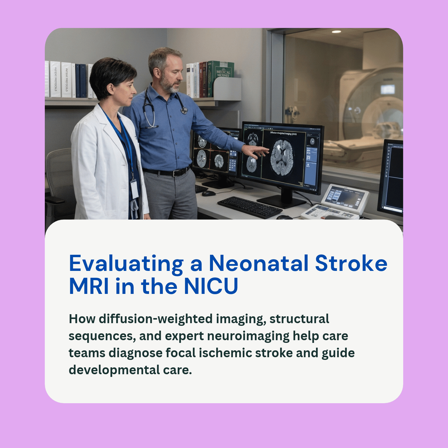
In cases of suspected focal injury, magnetic resonance imaging (MRI) serves as the definitive gold standard for neuroradiological evaluation. Ordering a neonatal stroke MRI allows care teams to visualize brain tissue without ionizing radiation, confirming whether an ischemic lesion or hemorrhagic event has occurred. However, an MRI scan is not a single instant answer or a predictor of exact future outcomes. Neuroradiologists and pediatric neurologists must combine advanced imaging sequences with physical neurological examinations and developmental follow-up. This educational guide explains what specialized MRI sequences reveal, why scan timing matters, how stroke evaluations differ from global injury scans, and how imaging fits into comprehensive care.
Key MRI Sequences and What They Reveal
A neonatal brain MRI protocol uses several specialized imaging sequences to evaluate blood vessels, brain tissue, and cellular swelling:
Diffusion-Weighted Imaging (DWI) and Apparent Diffusion Coefficient (ADC): DWI is the most sensitive sequence for detecting acute ischemic injury within hours of onset. When blood flow to a specific brain region is interrupted, cellular energy pumps fail, causing water molecules to shift inside brain cells (cytotoxic edema). DWI captures this restricted movement of water, making acute stroke tissue appear as a bright, distinct focal region on the screen.
Conventional T1-Weighted and T2-Weighted Imaging: These structural sequences provide detailed anatomical pictures. In the first few days following an ischemic event, conventional T1 and T2 sequences show localized tissue swelling. Over subsequent weeks and months, they help document structural remodeling, tissue reorganization, or focal cystic changes.
Magnetic Resonance Angiography (MRA) and Venography (MRV): MRA visualizes major arterial blood vessels in the brain to check for persistent blockages, vascular malformations, or arterial dissection. MRV evaluates the large venous sinuses to rule out sinovenous thrombosis, where a clot forms in the veins draining blood from the brain.
Timing of Imaging and Limitations of Early Findings
The timing of a neonatal stroke MRI heavily influences what neuroradiologists can see on the scan:
The First Week of Life (Days 1 to 7): DWI imaging achieves its highest sensitivity during the acute phase (typically between 24 hours and 7 days after the event). Obtaining an MRI scan during this early window clearly delineates the boundaries of the infarction and helps distinguish an acute stroke from older, antenatal brain changes.
Pseudonormalization (Days 7 to 14): As acute tissue swelling subsides during the second week after a stroke, the restricted diffusion signal on DWI gradually fades, a technical phenomenon known as pseudonormalization. If an MRI is performed exclusively during this window, an acute stroke lesion can temporarily appear less obvious on diffusion sequences, requiring careful reliance on structural T1 and T2 images.
Limitations of Scans Alone: While an MRI precisely locates where brain tissue was affected, early scans cannot accurately predict an individual child’s precise motor or cognitive development. The infant brain possesses remarkable neuroplasticity, meaning healthy surrounding tissue can frequently adapt and reorganize motor pathways over time. Furthermore, an MRI scan alone cannot establish the exact underlying cause of a blood clot or imply medical negligence.
Differentiating Focal Stroke Assessment from Other Imaging Protocols
Understanding how a focal stroke evaluation differs from other neonatal imaging assessments helps clarify the medical team’s diagnostic focus:
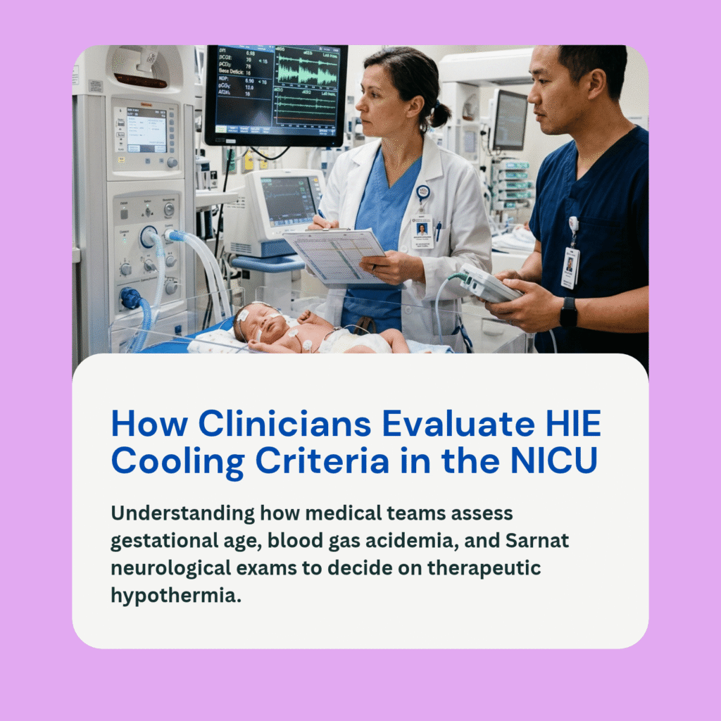
Focal Stroke Assessment vs. Global HIE Overview: Hypoxic-Ischemic Encephalopathy (HIE) involves systemic oxygen deprivation, producing widespread, symmetrical pattern changes in the basal ganglia, thalami, or watershed cortical regions. In contrast, a perinatal stroke evaluation focuses on localized, asymmetrical lesions confined to a single arterial territory (such as the left middle cerebral artery).
Focal Stroke Assessment vs. General Stroke Explainer: While general educational overviews explain what a stroke is, a specialized imaging evaluation focuses on the technical mechanics of neuroradiology: identifying arterial versus venous blockage, assessing vascular anatomy via MRA/MRV, and monitoring cellular water restriction on diffusion sequences.
Integrating Imaging with Examination and Follow-Up
An MRI scan provides an anatomical roadmap, but care decisions depend on how the infant functions:
Neonatologists and pediatric neurologists combine imaging findings with daily physical assessments of muscle tone, primitive reflexes, and spontaneous arm and leg movements (refer internal link: What Is a Newborn Neurological Exam After HIE?). Continuous brain wave monitoring (refer internal link: What Is Amplitude-Integrated EEG (aEEG) in the NICU?) ensures that electrographic seizures triggered by the stroke are managed effectively (refer internal link: How Are Neonatal Seizures Treated and Monitored in the NICU?).
Following discharge, families are connected with specialized developmental tracking through a high risk infant follow up clinic. Serial developmental assessments allow physical and occupational therapists to introduce targeted early intervention therapies as the child reaches key motor milestones.
Frequently Asked Questions (FAQ)
Does an infant need to be sedated to get a brain MRI?
In many neonatal units, doctors utilize a feed and wrap technique (also called feed and bundle). The baby is fed, swaddled comfortably, and allowed to fall asleep naturally before being placed securely in the MRI scanner with special ear protection, avoiding the need for general anesthesia.
Can an MRI show exactly when a neonatal stroke occurred?
Early MRI scans using diffusion-weighted imaging (DWI) can narrow the timing window to within a few days of birth. However, imaging cannot determine the exact minute or hour a blood clot formed, nor can it pinpoint the single underlying clinical cause.
If the MRI shows a stroke, does that mean my baby will have hemiplegia or cerebral palsy?
Not necessarily. While a stroke lesion in the motor cortex or internal capsule carries an increased risk for motor asymmetry, early physical and occupational therapy helps activate neuroplastic pathways. Many infants recover substantial motor function as they grow.
Is a neonatal stroke diagnosis on MRI proof of birth injury negligence?
No. Most perinatal arterial strokes occur due to complex, unpredictable vascular events, such as a clot originating in the placenta or temporary alteration in fetal blood flow. Establishing whether standard medical protocols were followed requires an independent clinical review of the entire obstetric, placental, and neonatal record.