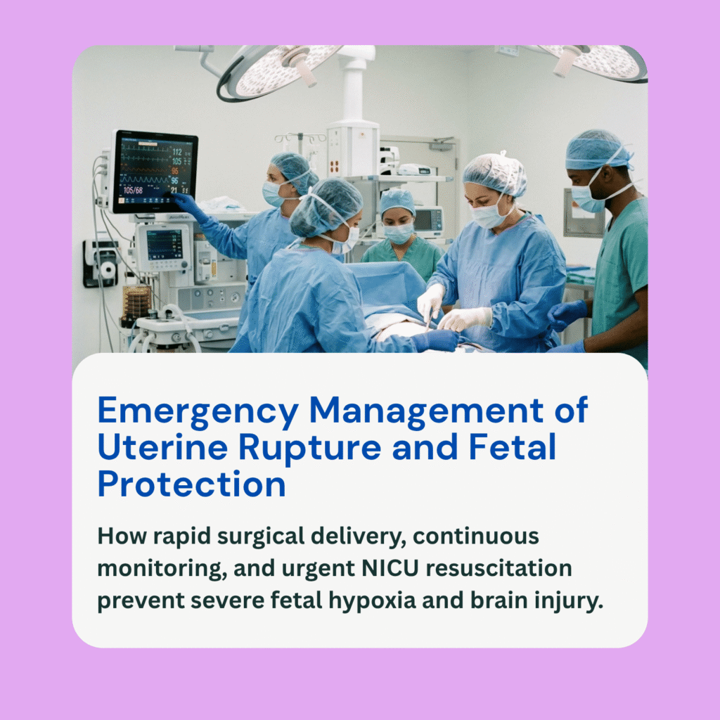
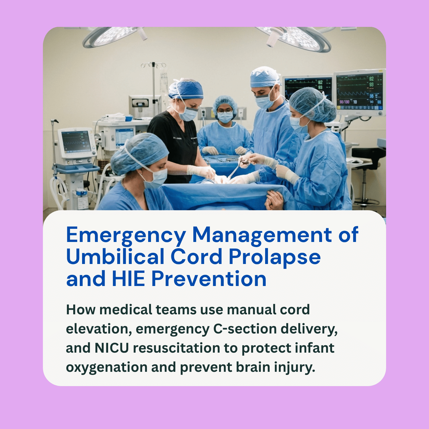

Can Umbilical Cord Prolapse Cause HIE or Brain Injury?
An umbilical cord prolapse is an urgent, life-threatening obstetric emergency that occurs when the umbilical cord slips down into the birth canal ahead of or alongside the baby’s presenting body part after the amniotic sac ruptures. Instead of floating freely in amniotic fluid, the umbilical cord becomes trapped between the baby’s body—usually the head or buttocks—and the mother’s bony pelvis or cervix.
Because the umbilical cord acts as the baby’s primary lifeline, transporting oxygenated blood from the placenta, physical pressure on the cord severely restricts or halts blood flow to the fetus. A primary concern for parents navigating this traumatic delivery experience is whether an umbilical cord prolapse HIE or permanent brain injury can occur. The answer is that a prolapsed cord presents an immediate risk of acute, severe fetal oxygen loss, which can cause Hypoxic-Ischemic Encephalopathy (HIE) and secondary brain damage if not relieved within minutes. However, neurological outcomes vary significantly based on how quickly the medical team identifies the prolapse and executes an emergency C-section. This guide explains the mechanisms behind cord prolapse, key risk factors, emergency warning signs, and management protocols.
Types of Umbilical Cord Prolapse and Risk Factors
Obstetricians categorize umbilical cord displacement into two primary forms based on physical presentation:
Overt Cord Prolapse: The umbilical cord slips completely past the baby’s presenting part and protrudes into the vagina or extends outside the labia after membranes rupture. This is an immediate, visible clinical emergency.
Occult Cord Prolapse: The umbilical cord sits alongside the presenting part within the birth canal but is not visible or palpable past the baby. While hidden, it remains subject to severe compression during labor contractions.
A cord prolapse occurs when the baby’s presenting part does not fully occupy or seal the lower uterine segment when the waters break. Several maternal and fetal risk factors increase this likelihood:
A primary risk factor is premature rupture of membranes (PROM) or preterm labor, where a smaller baby leaves extra space in the pelvic inlet. Polyhydramnios—an excessive volume of amniotic fluid—can trigger a sudden surge of fluid that flushes the umbilical cord downward when the amniotic sac ruptures. Malpresentation, such as a breech (feet or bottom first) or transverse (side-lying) baby, prevents the birth canal from being firmly plugged. Additional risk factors include carrying twins or higher-order multiples, having an abnormally long umbilical cord, grand multiparity, or medical interventions such as an artificial rupture of membranes (amniotomy) when the baby’s head is still high in the pelvis (unengaged).
Recognizing Warning Signs and Acute Oxygen Deprivation
Unlike gradual labor complications, a prolapsed umbilical cord presents abruptly and demands instant recognition from labor and delivery nurses and obstetricians:
Sudden, Prolonged Fetal Bradycardia: The most reliable electronic sign of a prolapse is an immediate, severe drop in fetal heart rate (prolonged decelerations or sustained bradycardia dropping below 100 or 80 beats per minute) immediately following the rupture of membranes.
Visible or Palpable Cord: Upon physical or vaginal examination following fluid release, clinicians may visually see the umbilical cord protruding from the vagina or feel a soft, pulsating cord ahead of the baby’s head.
Pathophysiology of Acute Brain Injury: As contractions push the baby downward against the trapped cord, blood flow through the umbilical vein and arteries drops to near zero. This produces acute systemic fetal hypoxemia and hypercapnia. Deprived of oxygen and glucose, the fetal brain enters acute cellular energy failure. Ion pumps across cell membranes collapse, triggering toxic intracellular swelling, membrane dissolution, and cellular tissue death. This process directly results in Hypoxic-Ischemic Encephalopathy (HIE).
Emergency Management and Medical Response
When an overt or occult cord prolapse occurs, time is measured in seconds. Medical guidelines emphasize a coordinated, rapid-response protocol to relieve physical pressure on the cord while preparing for surgical delivery:
Manual Cord Elevation: The single most critical immediate step is for a clinician to insert a gloved hand into the vagina and manually push the baby’s presenting head or body part upward off the umbilical cord. The clinician must hold this position continuously to relieve compression until the baby is delivered in the operating room.
Maternal Positioning and Bladder Filling: The mother is immediately placed in a position that uses gravity to shift the fetus toward the upper abdomen, such as the knee-chest position or severe Trendelenburg position (head lower than hips). In some cases, clinicians instill sterile saline into the mother’s urinary bladder via a catheter to gently lift the baby’s head off the cord.
Emergency C-Section and NICU Cooling: An emergency C-section must be executed as quickly as possible. Following delivery, the neonatal resuscitation team immediately stabilizes the baby’s airway, establishes mechanical ventilation, and evaluates acid-base levels via umbilical cord blood gas analysis. If the infant displays clinical signs of moderate to severe HIE within the first six hours of life, therapeutic hypothermia (cooling therapy) is initiated to slow metabolic stress and mitigate brain damage.
Neurological Outcomes and Long-Term Implications
The long-term impact of an umbilical cord prolapse depends on the total duration and severity of total oxygen deprivation before delivery is achieved:
When recognized instantly and managed with skilled manual elevation and rapid delivery, many babies recover fully without brain damage. However, if cord compression is prolonged, severe hypoxic brain injury can result in long-term neurological conditions. Potential outcomes include spastic or dyskinetic cerebral palsy, cognitive impairments, motor skill deficits, vision or hearing loss, and chronic seizure disorders.
Frequently Asked Questions (FAQ)
Can an umbilical cord prolapse be predicted or prevented during pregnancy?
An umbilical cord prolapse cannot be predicted with total certainty or completely prevented, as it happens suddenly when the amniotic sac breaks. However, doctors can identify high-risk situations—such as a breech presentation or polyhydramnios—and avoid elective amniotomy (breaking the water) when the baby’s head is unengaged.
How many minutes does a medical team have to deliver a baby during a cord prolapse?
While older guidelines referenced a 30-minute decision-to-delivery target for emergency C-sections, a total cord occlusion requires delivery as quickly as possible—ideally within 10 to 15 minutes of onset—to avoid irreversible brain damage or fetal death, unless manual elevation successfully maintains blood flow.
What is the difference between a cord prolapse and a nuchal cord?
A nuchal cord occurs when the umbilical cord is wrapped around the baby’s neck in the womb. A cord prolapse occurs when the cord slips ahead of the baby into the birth canal, where it becomes compressed between the baby’s body and the birth canal walls.
Can an umbilical cord prolapse birth injury involve medical negligence?
Yes. While the prolapse itself is often an unpreventable physical event, medical negligence can occur if providers perform an artificial rupture of membranes inappropriately on an unengaged baby, fail to recognize sudden fetal bradycardia, fail to manually elevate the baby off the cord, or cause unnecessary delays in executing an emergency C-section.