
Can Newborn Seizures Be a Sign of HIE?
A neonatal seizure is one of the most significant clinical indicators of neurological dysfunction in a newborn baby. Occurring most frequently within the first 24 to 48 hours after birth, seizures reflect abnormal, excessive electrical discharges across immature neural pathways in the brain. Unlike adult seizures, which often involve dramatic full-body convulsions, infant seizures can be remarkably subtle and easily overlooked without specialized monitoring.
A central question for parents in the Neonatal Intensive Care Unit (NICU) is whether newborn seizures HIE are connected. Hypoxic-Ischemic Encephalopathy (HIE)—a brain injury caused by severe oxygen deprivation and restricted blood flow around the time of birth—is the single leading cause of neonatal seizures, accounting for over 50% of all cases diagnosed in term infants. While seizures can also stem from metabolic imbalances, infections, or stroke, their onset in a newborn who experienced intrapartum distress serves as a critical diagnostic warning sign of hypoxic brain injury. This educational guide explores how infant seizures manifest, why they are often subtle, how medical teams diagnose them, and current treatment standards.
Why Newborn Seizures Manifest Differently and Subtle Warning Signs
The immature nervous system of a newborn baby processes electrical impulses differently than an adult or older child. Because the brain’s cerebral cortex is not fully myelinated, abnormal electrical discharges cannot easily spread across both hemispheres to produce generalized tonic-clonic convulsions. As a result, neonatal seizures frequently present as subtle physical behaviors:
Subtle Seizures: These are the most common type of seizures in newborns. They may appear as unusual ocular movements like rhythmic eye-blinking, persistent staring, or eyes rolling upward. Oral-buccal movements are also common, including repetitive smacking of the lips, rhythmic tongue thrusting, or unnatural sucking motions. Motor behaviors may resemble cycling movements of the legs (bicycled legs) or repetitive rowing movements of the arms.
Clonic Seizures: Characterized by rhythmic, slow jerking movements of a single limb, one side of the body, or specific facial muscles. These movements cannot be stopped by gently holding the baby’s flexing limb, which distinguishes a true clonic seizure from normal benign infant jitteriness.
Tonic Seizures: Marked by sustained, rigid extension or flexing of the arms, legs, or torso. These postural changes can mimic normal primitive reflexes or posturing but last longer and occur involuntarily.
Myoclonic Seizures: Present as sudden, rapid, single or repeated muscle twitches involving the arms, legs, or whole body.
The Pathophysiological Link Between HIE and Seizures
During an acute hypoxic event—such as cord prolapse, uterine rupture, or severe placental abruption—the infant’s brain is deprived of oxygen and glucose. Without these essential nutrients, the brain’s cellular energy pumps (sodium-potassium ATPase pumps) fail.
This energy failure destabilizes the cell membranes of neurons, causing an abnormal influx of sodium and calcium ions and an excessive buildup of the excitatory neurotransmitter glutamate. The resulting cellular hyperexcitability triggers continuous, disorganized electrical firing across brain tissue. This electrical dysregulation presents physically as seizures. Furthermore, active clinical seizures increase the brain’s metabolic demand and oxygen consumption, which can cause secondary brain tissue damage if the seizures are left unmanaged.
Diagnostic Evaluation: EEG, Imaging, and Lab Testing
Because many subtle physical behaviors mimic seizures and up to 80% of electrical seizures in neonates produce no visible physical symptoms (electroclinical dissociation), precise diagnostic testing is critical:
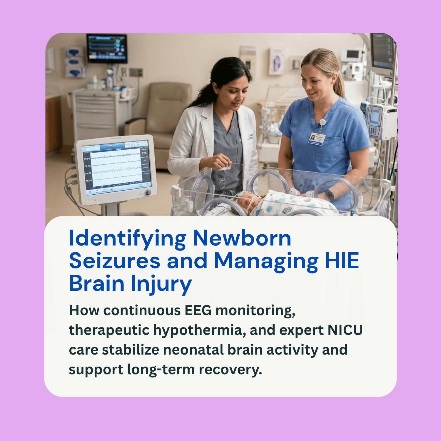
Amplitude-Integrated and Continuous Video EEG: Continuous Electroencephalography (EEG) combined with video recording is the gold standard for diagnosing neonatal seizures. In many NICUs, amplitude-integrated EEG (aEEG) is used at the bedside as a continuous monitoring tool. An EEG measures electrical waves in the brain, allowing neonatologists to confirm seizure activity, pinpoint where electrical spikes originate, and evaluate whether medical interventions are working.
Neuroimaging (MRI and Ultrasound): Magnetic Resonance Imaging (MRI) is the primary imaging modality used to evaluate the extent and pattern of brain injury caused by HIE. Brain MRIs—typically performed between 3 to 10 days of life—can identify restricted diffusion in key structures like the basal ganglia, thalamus, and white matter. Cranial ultrasound is also used at the bedside to rule out intraventricular hemorrhage or structural brain anomalies.
Laboratory Testing: Doctors conduct comprehensive blood panels to check blood glucose levels, ionized calcium, magnesium, electrolytes, arterial blood gases, and inflammatory markers, ruling out metabolic causes like severe hypoglycemia or active sepsis.
Medical Treatment, Therapeutic Hypothermia, and Follow-Up
Managing newborn seizures caused by HIE involves stabilizing brain metabolism, suppressing electrical storm activity, and providing long-term developmental tracking:
Therapeutic Hypothermia (Cooling Therapy): For infants born at or near term who show signs of moderate to severe HIE within six hours of birth, therapeutic hypothermia is the standard evidence-based medical treatment. The baby’s core body temperature is lowered to approximately 33.5°C (92.3°F) for 72 hours. Cooling therapy slows cellular metabolism, reduces inflammatory chemical cascades, limits secondary cell death, and significantly reduces the overall frequency and severity of neonatal seizures.
Antiseizure Medications: If clinical or electrical seizures persist, neonatologists administer intravenous antiseizure medications. Phenobarbital remains the primary first-line treatment, while second-line options include levetiracetam (Keppra) and fosphenytoin. Medical teams carefully adjust medication dosages using continuous EEG monitoring to achieve total electrographic seizure control.
Long-Term Follow-Up and Rehabilitation: Children who suffer seizures secondary to HIE require structured neurodevelopmental follow-up. A multidisciplinary care team—including pediatric neurologists, physical therapists, occupational therapists, and speech-language pathologists—monitors the child’s developmental milestones to identify and address any motor deficits, cognitive delays, cerebral palsy, or chronic epilepsy early in life.
Frequently Asked Questions (FAQ)
Are all newborn seizures caused by HIE?
No. While Hypoxic-Ischemic Encephalopathy (HIE) is the leading cause of neonatal seizures, other potential causes include severe neonatal hypoglycemia, central nervous system infections (such as meningitis or herpes simplex virus), stroke, congenital brain malformations, and metabolic disorders.
How can I tell the difference between normal infant jitteriness and a seizure?
Normal infant jitteriness usually consists of rapid, tremor-like movements triggered by sudden movement or noise, and the movement stops completely if you gently hold the baby’s arm or leg. Seizure movements are typically rhythmic, may involve subtle eye or lip movements, cannot be stopped by holding the limb, and can occur spontaneously while the baby is resting.
Do newborn seizures mean my baby will have epilepsy forever?
Not necessarily. Many infants who experience seizures due to acute HIE only have seizures during the acute phase of brain injury in the NICU. Once the initial injury stabilizes and healing occurs, many babies can be safely weaned off antiseizure medications without developing chronic epilepsy, though long-term neurological monitoring is essential.
Can delay in treating newborn seizures lead to medical malpractice concerns?
While HIE itself stems from perinatal stress, failing to recognize subtle seizure signs, delaying continuous EEG monitoring, failing to offer cooling therapy within the critical six-hour window, or leaving clinical seizures untreated can worsen secondary brain injury and may warrant legal review.