
What Is a Newborn Neurological Exam After HIE?
When a newborn experiences Hypoxic-Ischemic Encephalopathy (HIE)—a brain injury resulting from oxygen deprivation and restricted blood flow near the time of delivery—medical teams must continually assess how well the infant’s central nervous system is functioning. While bedside monitors track vital signs and imaging scans provide structural pictures of brain tissue, the newborn neurological examination provides direct, real-time functional information about the baby’s neurological state.
For parents in the Neonatal Intensive Care Unit (NICU), watching pediatric neurologists and neonatologists perform a newborn neurological exam HIE evaluation can raise many questions. Healthcare providers use structured, physical assessments to detect signs of neonatal encephalopathy, determine whether therapeutic cooling therapy is needed, and track neurological recovery over time. However, a single neurological exam is not a crystal ball for an individual child’s future. Instead, serial physical examinations serve as a foundational clinical baseline that must be integrated with continuous amplitude-integrated EEG (aEEG), umbilical cord blood gases, and diagnostic brain MRI scans. This guide explains what the exam measures, how structured systems like Sarnat staging work, and how clinical findings guide ongoing NICU care.
Core Components of the Neonatal Neurological Assessment
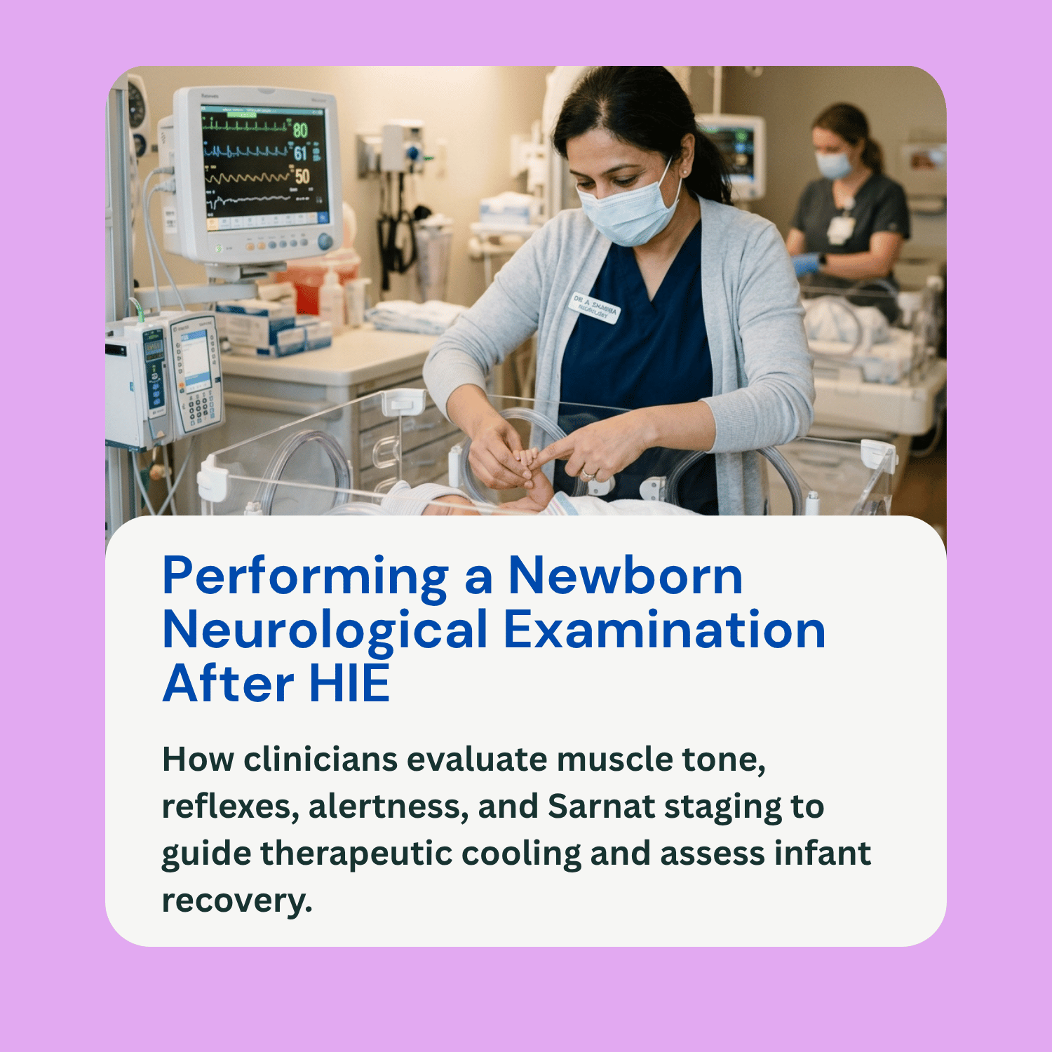
A newborn neurological examination is a gentle, systematic physical evaluation designed to check primitive reflexes, brainstem integrity, and motor pathways. Clinicians evaluate several key clinical domains:
Level of Consciousness and Alertness: The examiner observes whether the baby is normally active, hyperalert (excessively wide-eyed or irritable), lethargic (difficult to wake), or stuporous/comatose. Altered alertness is often one of the earliest signs of acute cerebral dysfunction.
Muscle Tone and Posture: The physician assesses both resting posture and active muscle tone. Normal term infants keep their arms and legs flexed near their chest. In HIE, babies may exhibit hypotonia (floppiness or severe weakness) or hypertonia (rigid, stiff extension of the limbs). Muscle tone is checked through passive movement of the arms and legs, as well as tests like head lag when pulled to sit.
Spontaneous Movement: Clinicians observe the quality, symmetry, and frequency of the baby’s natural movements. Smooth, symmetrical arm and leg movements indicate functional motor pathways, whereas absent movements, asymmetrical responses, or involuntary rhythmic twitches may indicate motor deficits or electrographic seizure activity.
Primitive and Brainstem Reflexes: The exam checks essential newborn reflexes that depend on intact deep brain and spinal pathways:
- Suck and Swallow Reflexes: Measures the infant’s ability to coordinate feeding and protect their airway.
- Moro (Startle) Reflex: Evaluates how the baby responds to a sudden change in position; a weak or absent Moro reflex signals nervous system depression.
- Grasp Reflexes: Checks the strength of the baby’s automatic grip when a finger is placed in their palm or sole.
- Pupillary and Oculomotor Reflexes: Tests eye movement and pupil constriction in response to light to evaluate brainstem function.
Categorizing Encephalopathy: The Sarnat Staging System
To standardize findings across different medical providers, neonatologists frequently utilize the Sarnat Staging System, which classifies neonatal encephalopathy into three clinical stages:
Mild Encephalopathy (Sarnat Stage 1): Characterized by hyperalertness, uninhibited sympathetic responses (such as dilated pupils or rapid heart rate), jitteriness, and slightly increased muscle tone. Seizures are absent, and full recovery typically occurs within a few days.
Moderate Encephalopathy (Sarnat Stage 2): Marked by lethargy, hypotonia (weak muscle tone), suppressed primitive reflexes (weak suck and Moro), and constricted pupils. Electrographic seizures frequently occur in Stage 2. Moderate encephalopathy is a primary clinical indicator for initiating 72-hour therapeutic hypothermia (cooling therapy) to protect brain tissue.
Severe Encephalopathy (Sarnat Stage 3): Represents severe neurological depression. The newborn is stuporous or comatose, completely limp (flaccid), with absent reflexes, non-reactive pupils, and suppressed breathing effort requiring mechanical ventilation. Seizures may be present or suppressed by severe cellular dysfunction.
Why Serial Examinations and Integrated Data Matter
A single neurological examination represents a moment-in-time snapshot. Because the newborn brain is dynamic and rapidly evolving following a hypoxic insult, clinicians rely on repeated, serial examinations performed over hours and days:
Tracking Clinical Trajectory: Serial examinations show whether an infant’s neurological status is improving, remaining stable, or deteriorating. A transition from moderate lethargy back to normal tone and robust feeding during rewarming after cooling therapy provides a positive clinical sign.
Combining Physical Exams with Objective Diagnostics: Physical findings are always interpreted alongside objective diagnostic data:
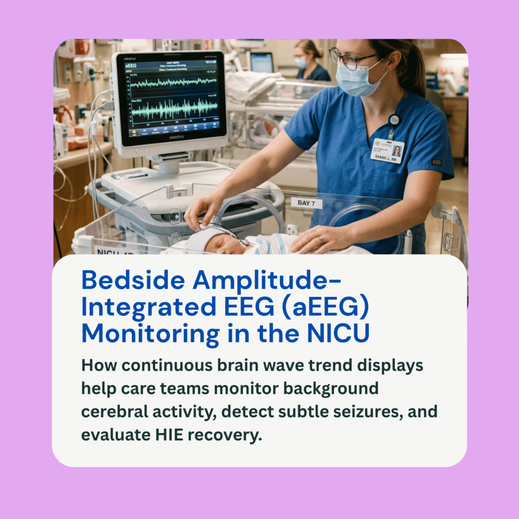
- Continuous aEEG and Video EEG: Physical exams detect visible muscle movements, while continuous EEG monitors detect silent electrographic seizures that occur without physical signs.
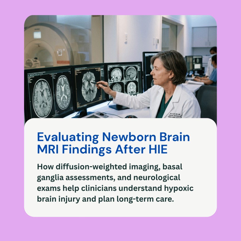
- Neonatal Brain MRI: While an MRI reveals structural cellular injury (such as basal ganglia or watershed lesions), the physical neurological exam shows how those structural findings manifest in actual motor function and alertness.
Focusing on Developmental Potential: Because infant brains possess significant neuroplasticity, clinicians avoid predicting an individual child’s ultimate outcome based solely on initial NICU exams. Instead, physical exam findings guide long-term early intervention referrals, such as pediatric physical, occupational, and speech therapies.
Frequently Asked Questions (FAQ)
Is a newborn neurological exam painful for the baby?
No. The examination consists entirely of gentle physical maneuvers, light tactile stimulation, visual observation, and checking natural reflexes. Care providers perform the exam carefully to avoid overstimulating an infant undergoing recovery or cooling therapy.
How does a doctor determine if a baby needs cooling therapy based on the neurological exam?
Clinicians perform a structured neurological exam within the first six hours of life. If the infant displays signs of moderate or severe encephalopathy (such as lethargy, hypotonia, and absent reflexes) alongside laboratory evidence of metabolic acidemia, therapeutic hypothermia is started immediately.
Can a baby with abnormal muscle tone in the NICU recover normal movement later?
Yes. Abnormal muscle tone (hypotonia or hypertonia) during the acute phase of HIE reflects temporary cerebral shock and cellular swelling. As brain swelling subsides and therapeutic interventions take effect, many infants show significant improvement in muscle tone and motor function.
How do neurological exam records factor into birth injury reviews?
In medical-legal reviews, serial neurological exam notes provide crucial timeline evidence. They document exactly when signs of encephalopathy first appeared after birth, how severe the neurological depression was, and whether the clinical team recognized moderate-to-severe HIE in time to initiate cooling therapy within the required 6-hour window.