
What Can a Newborn Brain MRI Show After HIE?
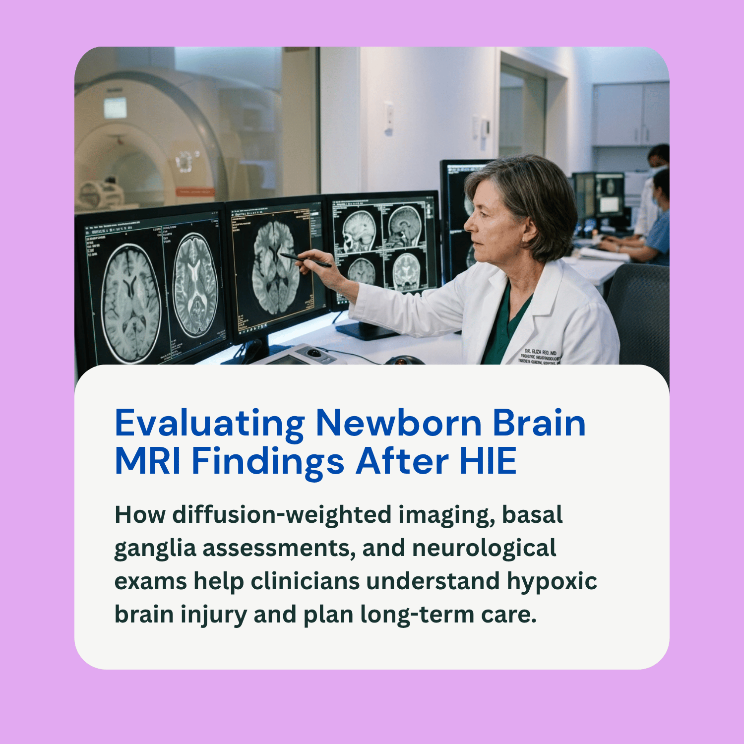
When a newborn experiences Hypoxic-Ischemic Encephalopathy (HIE)—a brain injury caused by oxygen deprivation and restricted blood flow near the time of birth—neonatologists rely on specialized neuroimaging to assess the brain’s structural integrity. Magnetic Resonance Imaging (MRI) is the single most informative and sensitive diagnostic tool for evaluating the presence, location, and extent of hypoxic-ischemic brain damage in infants.
For parents in the Neonatal Intensive Care Unit (NICU), reviewing a newborn brain MRI HIE report can feel daunting. An MRI provides detailed visual evidence of which brain structures were affected by oxygen starvation and helps clinicians understand the timing and mechanism of the injury. However, an MRI scan is not a crystal ball. Imaging results represent a static picture that must be interpreted alongside continuous bedside EEG monitoring, physical neurological examinations, and long-term developmental assessments. This educational guide explains why and when brain MRIs are performed, common imaging patterns, diagnostic limitations, and how results fit into overall care planning.
Timing and Imaging Techniques in Neonatal MRI
Timing is critical when scheduling a brain MRI for a newborn with suspected or confirmed HIE, as cellular changes evolve over the days following an acute hypoxic insult:
Optimal Imaging Window: For infants who receive therapeutic hypothermia (cooling therapy), the primary diagnostic MRI is typically performed between 3 to 10 days of life, usually after the 72-hour cooling and rewarming protocols are completed. Scanning during this window provides the clearest picture of restricted diffusion and acute cellular injury. Scanning too early (within the first 24 to 48 hours) may underestimate the extent of brain tissue damage because secondary injury processes are still evolving.
Diffusion-Weighted Imaging (DWI): Standard anatomical MRI sequences (such as T1-weighted and T2-weighted images) capture detailed structural views. However, Diffusion-Weighted Imaging (DWI) and calculated Apparent Diffusion Coefficient (ADC) maps are particularly critical during the acute phase. DWI detects subtle cytotoxic edema—the cellular swelling that occurs immediately after brain cells suffer energy failure—allowing radiologists to spot hypoxic injury days before it becomes visible on conventional MRI sequences.
Magnetic Resonance Spectroscopy (MRS): In many tertiary NICUs, doctors also perform MR Spectroscopy during the same MRI session. MRS measures biochemical markers in brain tissue, evaluating levels of N-acetylaspartate (NAA), lactate, and choline. A sudden spike in lactate combined with reduced NAA signals metabolic crisis and cell injury.
Common Injury Patterns Identified on HIE Brain MRIs
Depending on whether the oxygen loss was acute and profound or partial and prolonged, hypoxic-ischemic injury typically affects specific deep structures or peripheral regions of the newborn brain:
Deep Grey Matter Pattern (Basal Ganglia and Thalamus): This pattern commonly results from an acute, profound hypoxic event—such as an umbilical cord prolapse, severe placental abruption, or uterine rupture. The basal ganglia and thalamus are deep brain structures with extremely high metabolic demands. On MRI, acute injury to these regions appears as hyperintensity (bright signals) on T1-weighted images or restricted diffusion on DWI, often involving the posterior limb of the internal capsule (PLIC). Injury to the basal ganglia and thalamus strongly correlates with future motor impairments, including cerebral palsy.
Watershed / Parasagittal Pattern: This pattern typically occurs during partial, prolonged oxygen starvation, where blood pressure drops gradually. The brain prioritizes blood flow to its metabolic core, leaving the outer “watershed” boundaries between major cerebral arteries vulnerable. MRI scans in these cases show injury along the parasagittal white matter and cortex, which may influence cognitive development, language processing, and spatial orientation.
Total / Diffuse Cerebral Injury: In severe, catastrophic hypoxic events, imaging may show widespread involvement across both deep grey matter and cortical white matter.
Limitations of Imaging and the Complete Clinical Picture
While a newborn brain MRI provides essential structural data, medical guidelines emphasize that imaging alone cannot precisely predict a child’s exact long-term functional outcome:
Neuroplasticity and Individual Variability: The infant brain possesses remarkable neuroplasticity—the ability to rewire connections and compensate for damaged areas over time. Two infants with similar MRI findings may demonstrate significantly different functional abilities as they grow.
Correlating with Neurological Exams: A brain MRI must always be evaluated in tandem with serial physical examinations performed by a pediatric neurologist. A child’s clinical presentation—such as muscle tone (hypotonia vs. hypertonia), primitive reflexes, feeding capabilities, and alertness—provides immediate functional information that an MRI scan cannot capture.
Role of Continuous EEG: Amplitude-integrated and continuous video EEG monitoring tracks functional brain wave activity in real time, detecting subtle electrographic seizures that MRI scans cannot measure.
Long-Term Follow-Up: Comprehensive neurodevelopmental tracking through early intervention programs, physical therapy assessments, and regular pediatric neurology visits provides the true measure of a child’s developmental trajectory.
Frequently Asked Questions (FAQ)
Is an MRI safe for a newborn in the NICU?
Yes. An MRI uses powerful magnetic fields and radio waves, not ionizing radiation (like X-rays or CT scans), making it very safe for newborns. NICU transport teams carefully monitor the infant’s vital signs and temperature throughout the procedure.
Why did my baby have a cranial ultrasound before getting an MRI?
Cranial ultrasound is a fast, portable test performed right at the bedside to check for major brain bleeding (intraventricular hemorrhage) or structural abnormalities. However, ultrasound cannot clearly visualize early cytotoxic edema or deep grey matter injury, which is why an MRI is necessary later.
Can a normal brain MRI guarantee that my baby will not have developmental delays?
A completely normal brain MRI following suspected HIE is highly reassuring and indicates that severe structural injury did not occur. However, it cannot guarantee the absolute absence of mild learning differences or subtle developmental delays later in childhood, which is why ongoing neurodevelopmental follow-up remains important.
How do brain MRI findings factor into legal reviews for birth injuries? In legal evaluations, a brain MRI provides objective timing evidence. Specific injury patterns—such as acute basal ganglia lesions—help expert neonatologists determine whether oxygen starvation occurred during active labor, which is then evaluated against electronic fetal heart rate monitoring strips to assess clinical standard of care.