
What Is Amplitude-Integrated EEG (aEEG) in the NICU?
When a newborn is admitted to the Neonatal Intensive Care Unit (NICU) following a complicated delivery, birth trauma, or suspected Hypoxic-Ischemic Encephalopathy (HIE), monitoring brain function becomes a top medical priority. While heart rate, respiratory rate, and blood pressure monitors are standard bedside fixtures, evaluating real-time cerebral activity requires specialized equipment.
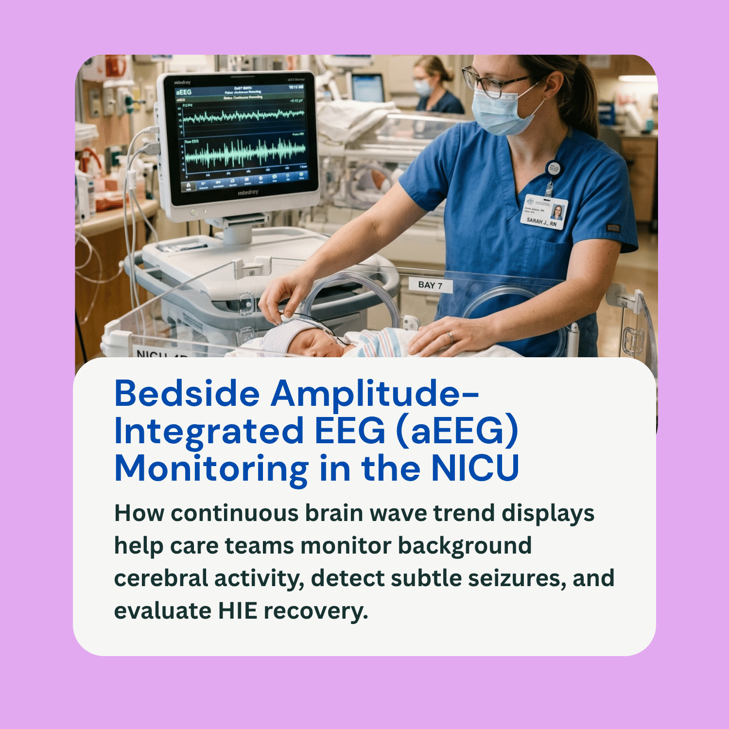
One of the primary tools neonatologists use at the bedside is amplitude-integrated electroencephalography (aEEG). An amplitude integrated EEG newborn monitor provides care teams with a simplified, continuous, long-term trend display of the baby’s electrical brain activity. By converting raw brain wave signals into an easy-to-read bedside trace, an aEEG allows nurses and neonatologists to detect subtle electrical changes, identify silent seizures, and track brain function over hours or days. However, an aEEG trace is not a standalone diagnostic test. It works as a continuous screening tool that must be interpreted alongside physical neurological examinations, conventional multi-channel EEGs, and brain MRI imaging. This educational guide explains how aEEG operates, why NICUs rely on it, how it differs from traditional EEG, and its role in comprehensive care.
How aEEG Works and Why NICU Teams Use It
An aEEG machine measures the background electrical activity generated by cerebral cortical neurons. Small, non-invasive sensors or hydrogel electrodes are placed on the infant’s scalp, typically using a reduced number of channels (such as two to four sensors across the parietal and central regions of the head).
The device records the raw brain wave signal, filters out low- and high-frequency noise, compresses the amplitude of the electrical signals logarithmically, and displays the activity as a continuous, slowly moving trend on a bedside screen.
NICU teams utilize aEEG continuous monitoring for several critical clinical reasons:
Assessing Background Brain Function: An aEEG displays baseline cerebral electrical activity, helping clinicians determine whether a newborn’s brain function is normal (continuous background activity), moderately abnormal (discontinuous activity), or severely depressed (low voltage or flat-line suppression).
Identifying Subclinical (Silent) Seizures: Up to 80% of newborn seizures after HIE are subclinical, meaning they produce no visible physical twitching, jerking, or eye movements. An aEEG alerts nurses to sudden spikes in background electrical amplitude that signal electrographic seizures, prompting immediate medical response.
Guiding Therapeutic Hypothermia: During cooling therapy (therapeutic hypothermia) for HIE, aEEG provides continuous feedback on whether cooling is successfully stabilizing brain activity and suppressing abnormal electrical firing.
Tracking Sleep-Wake Cycling: As a newborn recovers from an acute hypoxic event, the return of regular sleep-wake cycling on the aEEG trace serves as a positive clinical indicator of neurological recovery.
Key Differences: aEEG vs. Conventional EEG
While both tests record electrical brain signals, aEEG and conventional diagnostic EEGs serve complementary, distinct roles in the NICU environment:
Channel Count and Coverage: A conventional EEG uses 16 to 20 or more scalp electrodes placed in a full international 10–20 system montage. This provides high-resolution coverage across all regions of the brain. An aEEG uses only 2 to 4 electrodes, focusing primarily on central brain regions.
Interpretation Speed: A conventional EEG requires real-time review by a trained pediatric neurologist or neurophysiologist to read intricate, fast-moving wave lines. An aEEG compresses time (typically displaying 6 centimeters of trend data per hour), allowing NICU nurses and neonatologists to visually assess hours of trended brain activity at a glance at the bedside.
Availability and Continuity: Conventional EEGs are usually recorded in shorter diagnostic sessions (e.g., 30 to 60 minutes) when a neurologist is available to set up and review the trace. In contrast, an aEEG can be initiated rapidly by NICU staff and run continuously for 72 hours or longer throughout cooling therapy.
Limitations of aEEG and the Complete Clinical Picture
While aEEG is an invaluable bedside tool, medical guidelines emphasize its inherent technical limitations:
Risk of Artifact Interference: Because aEEG compresses electrical signals, muscle twitches, scalp movement, chest physiotherapy, and electrical noise from surrounding NICU equipment can occasionally mimic seizure spikes or distort background readings.
Detecting Focal or Short Seizures: Because aEEG uses few scalp electrodes, brief electrographic seizures (lasting under 30 seconds) or seizures originating in deep or isolated regions of the brain may not appear on the compressed trend line.
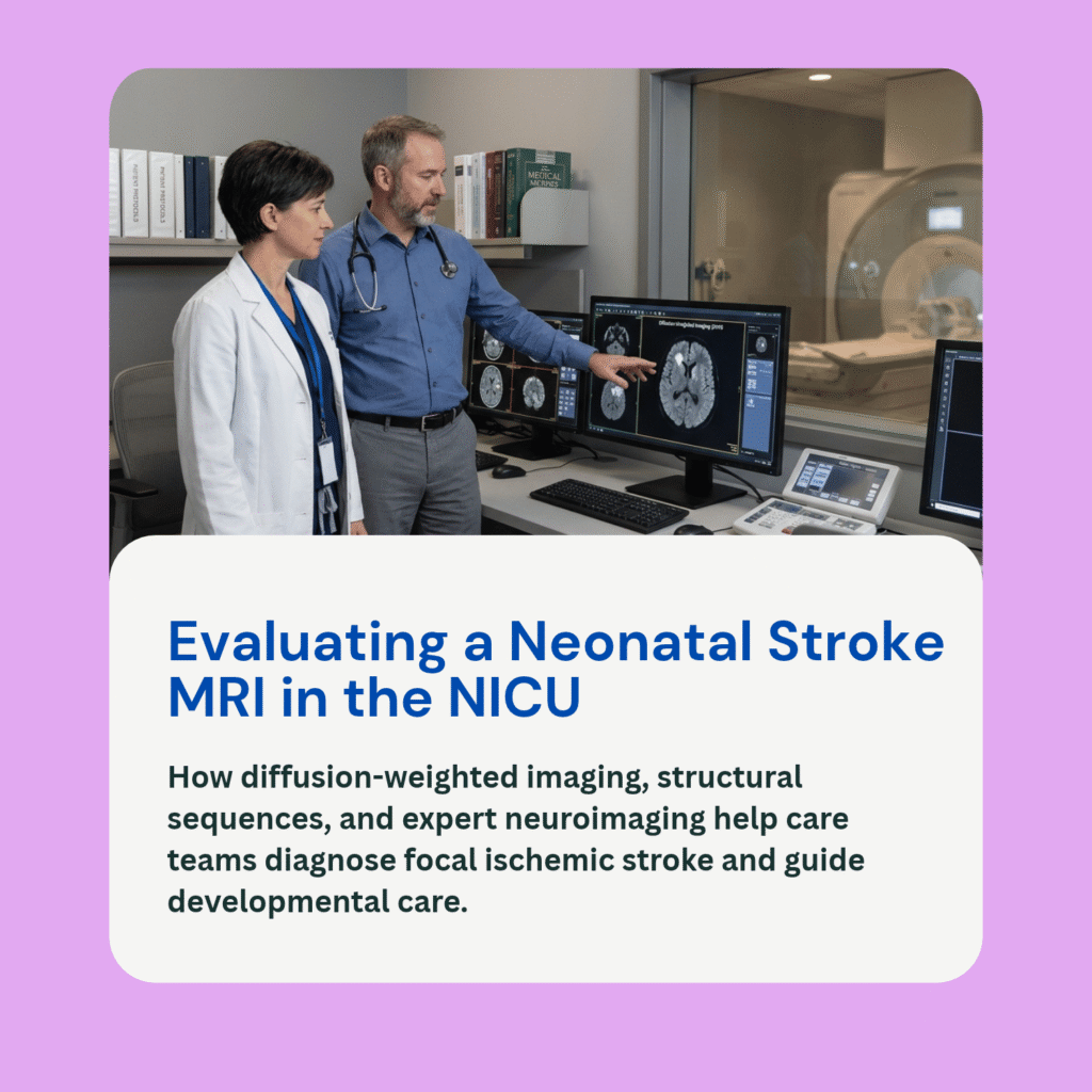
Integrating Full Diagnostic Data: Neonatologists never base clinical decisions or outcome predictions on an aEEG trace alone. An abnormal aEEG pattern prompts a request for a formal, multi-channel conventional EEG to confirm seizure activity and map specific focus areas. Furthermore, the aEEG trend is always correlated with daily neurological physical exams, arterial cord blood gas histories, metabolic labs, and high-resolution neonatal brain MRI scans performed between 3 to 10 days of life.
Frequently Asked Questions (FAQ)
Does putting an aEEG on my baby cause any pain?
No. Placing an aEEG involves applying soft hydrogel stickers or tiny needle electrodes gently onto the infant’s scalp. The monitoring process is completely non-invasive and painless.
Can an aEEG tell if my baby will have long-term developmental delays?
An aEEG provides valuable real-time information about acute brain function and recovery during the first days of life. While persistently flat or severely depressed background patterns are associated with higher neurological risk, an aEEG alone cannot predict an individual child’s precise long-term developmental path.
How long does a baby stay on an aEEG in the NICU?
For infants undergoing therapeutic hypothermia for HIE, aEEG monitoring typically continues throughout the entire 72-hour cooling phase and often extends through the rewarming period to ensure electrical stability.
How do aEEG records factor into medical malpractice reviews?
In birth injury reviews, continuous aEEG strips provide an objective timeline of when electrical brain dysfunction or seizures began. Experts evaluate whether the care team recognized abnormal background suppression or seizure spikes promptly and initiated appropriate antiseizure treatment or therapeutic hypothermia without delay.