
What Are Late Decelerations During Labor?
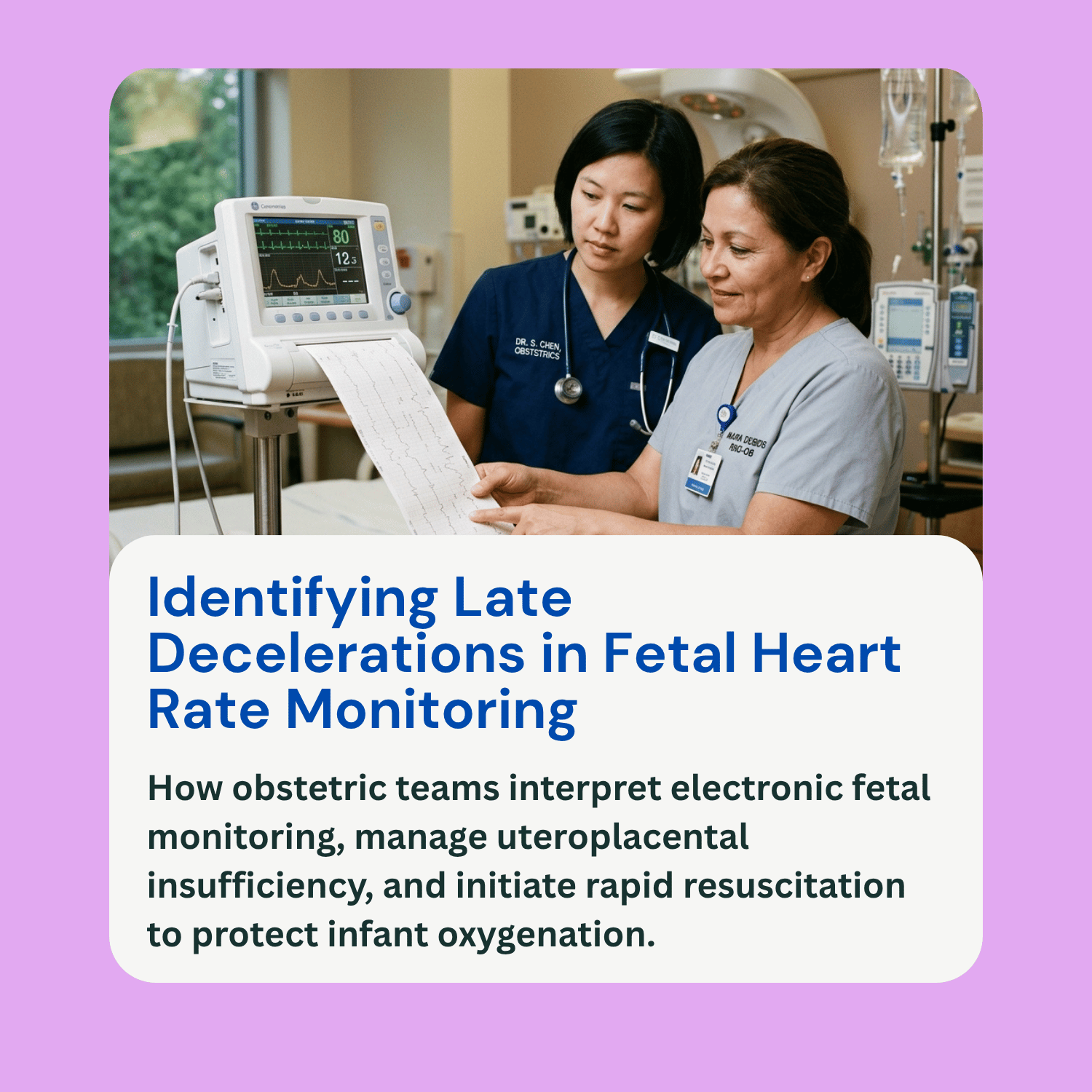
During labor and delivery, healthcare providers continuously track the baby’s health using electronic fetal monitoring (EFM). This monitoring system produces a continuous visual readout showing two primary measurements: the mother’s uterine contractions and the baseline fetal heart rate. Evaluating how the baby’s heart rate reacts during and immediately following contractions provides crucial insight into how well the fetus is tolerating labor.
One of the most concerning findings on a fetal heart trace is a pattern known as late decelerations fetal heart rate drops. A late deceleration is a smooth, gradual decrease in the baby’s heart rate that begins after a uterine contraction has already started, reaches its lowest point (nadir) after the peak of the contraction, and slowly returns to baseline only after the contraction has fully ended. This specific timing indicates that the fetus is experiencing reduced blood flow and oxygen exchange during contractions. This guide explains the causes of late decelerations, how they differ from other heart rate changes, how medical teams evaluate recurring patterns, and why a rapid clinical response is essential.
Comparing Fetal Heart Rate Deceleration Patterns
To accurately evaluate fetal well-being, clinicians categorize heart rate dips into three main types based on their shape, timing, and relationship to uterine contractions:
Early Decelerations: These are benign, symmetrical dips in the fetal heart rate that mirror the exact shape and timing of a contraction. The lowest point of the heart rate dip aligns precisely with the peak of the contraction. Early decelerations are caused by temporary head compression as the baby moves through the birth canal, which stimulates the vagus nerve. They are considered a normal physical response to labor and do not indicate fetal distress or hypoxia.
Variable Decelerations: These are abrupt drops in fetal heart rate where the speed of the decline is rapid, dropping sharply in less than 30 seconds. They vary significantly in duration, depth, and timing relative to contractions, often resembling a distinct “V” or “W” shape on the monitoring strip. Variable decelerations are primarily caused by umbilical cord compression, such as when the cord is wrapped around the baby’s neck (nuchal cord) or squeezed during a contraction. While occasional mild variables are common, persistent or deep variable decelerations require intrapartum management.
Late Decelerations: Unlike early or variable decelerations, late decelerations feature a gradual, subtle drop in heart rate where the nadir occurs well after the peak of the contraction. The key distinguishing factor is this delayed timing. Even if a late deceleration is shallow—dropping by only 5 to 15 beats per minute below baseline—its persistent late alignment signals underlying uteroplacental insufficiency and demands immediate clinical attention.
Causes of Uteroplacental Insufficiency and Late Decelerations
A late deceleration is a direct physiological response to transient fetal hypoxia. During a normal contraction, blood flow through the uterine muscle temporarily stops, forcing the fetus to rely on the reserve of oxygen stored in the placenta. If the placenta is healthy and maternal blood flow is sufficient, the fetus tolerates this brief pause easily. However, if the placenta is compromised or maternal blood pressure drops, the fetus suffers an acute drop in oxygen levels (hypoxemia), triggering a chemoreceptor-mediated response that slows the heart rate.
Several underlying conditions and labor factors can cause or worsen uteroplacental insufficiency:
Maternal hypotension is a frequent cause, often occurring as a side effect following epidural anesthesia placement, which relaxes blood vessels and reduces blood flow to the uterus. Uterine tachysystole—defined as more than five contractions in a ten-minute period—leaves insufficient rest time between contractions, rapidly depleting placental oxygen reserves. This condition is often caused by excessive administration of labor-inducing medications like oxytocin (Pitocin). Maternal medical conditions such as severe preeclampsia, chronic hypertension, gestational diabetes, or vascular disease also impair placental perfusion. Furthermore, post-term pregnancies, placental abruption, or structural placental failure reduce the functional tissue available for gas exchange.
How Clinicians Evaluate Recurring Patterns and Categorize Risk
Obstetricians and labor nurses evaluate late decelerations within the context of the overall fetal heart rate strip using a standardized three-tier classification system established by the American College of Obstetricians and Gynecologists (ACOG):
Category I (Normal): A Category I strip shows a stable baseline heart rate between 110 and 160 beats per minute, moderate baseline variability, and no late or variable decelerations. This indicates normal fetal acid-base status and requires standard routine monitoring.
Category II (Indeterminate): This broad category includes fetal heart rate patterns that require careful evaluation and continued surveillance. Occasional or intermittent late decelerations accompanied by moderate baseline variability fall into Category II. While not immediately definitive of severe birth asphyxia, these patterns signal that the baby is utilizing its physiological reserves and requires active intervention to prevent further deterioration.
Category III (Abnormal): A Category III strip is a clinical emergency indicating abnormal fetal acid-base balance and severe hypoxia. It is defined by persistent, recurrent late decelerations occurring with absent baseline variability, severe bradycardia, or a sinusoidal pattern. Category III patterns demand urgent resuscitation and prompt delivery.
Why Timely Clinical Response Matters
Recognizing late decelerations early allows the medical team to initiate intrauterine resuscitation maneuvers aimed at improving blood flow and oxygen delivery to the fetus. Standard first-line resuscitation protocols include changing the mother’s position to her left side to relieve pressure on the inferior vena cava, administering an intravenous fluid bolus to raise maternal blood pressure, providing supplemental oxygen if indicated, and stopping or reducing oxytocin infusions to slow down contractions.
If conservative resuscitation measures fail to eliminate recurrent late decelerations, or if the fetal heart rate trace progresses to a Category III pattern showing absent variability, immediate action is required. Prolonged, uncorrected uteroplacental insufficiency deprives the fetal brain of oxygen, leading to metabolic acidosis, hypoxic-ischemic encephalopathy (HIE), cerebral palsy, and long-term neurological injury. Timely decision-making—including expediting a C-section or operative vaginal delivery—is critical to preventing irreversible birth trauma.
Frequently Asked Questions (FAQ)
Are late decelerations always a sign of permanent fetal damage?
No. An isolated or brief period of late decelerations that responds quickly to intrauterine resuscitation maneuvers does not mean the baby will suffer brain damage. However, persistent, uncorrected late decelerations over a prolonged period indicate progressive oxygen deprivation, which increases the risk of hypoxic injury.
What is the difference between late decelerations and variable decelerations?
Late decelerations are smooth, gradual drops in heart rate caused by reduced blood and oxygen flow through the placenta, occurring after the peak of a contraction. Variable decelerations are abrupt, sharp drops in heart rate caused by physical compression of the umbilical cord, occurring at varying times relative to contractions.
How does epidural anesthesia cause late decelerations?
Epidural anesthesia can cause a sudden drop in the mother’s blood pressure (hypotension). When maternal blood pressure drops, less blood reaches the placenta during contractions, reducing oxygen delivery to the fetus and triggering late decelerations.
Can medical errors contribute to severe late decelerations?
Yes. Over-administering oxytocin (Pitocin) can cause uterine tachysystole, forcing contractions too close together and causing late decelerations. Additionally, failing to recognize late decelerations on the fetal monitor, delaying intrauterine resuscitation, or delaying an emergency C-section when a Category III pattern appears can constitute medical negligence.