
What Is a Subgaleal Hemorrhage in a Newborn?
A subgaleal hemorrhage newborn diagnosis represents one of the most critical neurological emergencies that can occur during labor and delivery. This condition develops when emissary veins—which connect the venous sinuses inside the skull to the scalp veins—rupture, causing blood to pool in the subgaleal space. This loose connective tissue layer sits between the periosteum (the membrane covering the skull bones) and the epicranial aponeurosis (the fibrous sheet of the scalp).
Because the subgaleal space spans across the entire cranial vault without being bound by skull suture lines, it forms a vast anatomical reservoir. In severe cases, an infant can lose 20% to 40% or more of their total blood volume into this hidden space, making rapid recognition and immediate intervention essential to prevent life-threatening complications.
Causes and Key Risk Factors
A subgaleal hemorrhage is almost always caused by mechanical traction and shear forces applied to the baby’s scalp during a difficult delivery, which tear delicate emissary blood vessels. While it can occasionally occur during an unassisted vaginal birth, key maternal and delivery risk factors significantly increase the odds.
Assisted delivery tools represent the primary cause of subgaleal hemorrhages, with approximately 90% of cases linked to vacuum extraction. When a vacuum extractor cup or forceps are applied with excessive force, pulled repeatedly, placed incorrectly on the infant’s head, or left on for extended periods, the scalp tissue is pulled away from the underlying skull, severing emissary veins. Other notable risk factors include a prolonged second stage of labor, fetal head malpositioning (such as occiput posterior), macrosomia (large birth weight), cephalopelvic disproportion (CPD), and primiparity (first-time mothers).
Recognizing Symptoms of Subgaleal Hemorrhage
Unlike localized, bounded head swelling like a cephalohematoma, a subgaleal hemorrhage spreads across the head and can expand rapidly. Because bleeding occurs underneath a wide tissue plane, physical signs and symptoms of internal blood loss may appear hours after delivery:
Diffusely Expanding “Boggy” Scalp Swelling: A signature sign is progressive, fluid-like (fluctuant) swelling over the infant’s head that crosses suture lines. The swelling often shifts with gravity, pushes the baby’s ears forward, or extends down toward the neck and forehead.
Signs of Hypovolemic Shock: As significant blood volume pools in the scalp, the newborn develops rapid heart rate (tachycardia), low blood pressure (hypotension), pale or mottled skin tone (pallor), cold extremities, and weak peripheral pulses.
Neurological Deterioration and Distress: Deprived of adequate circulating oxygen and blood volume, the infant may demonstrate severe lethargy, poor muscle tone (hypotonia), respiratory distress, difficulty feeding, or seizures.
Diagnosis and Emergency Medical Treatment
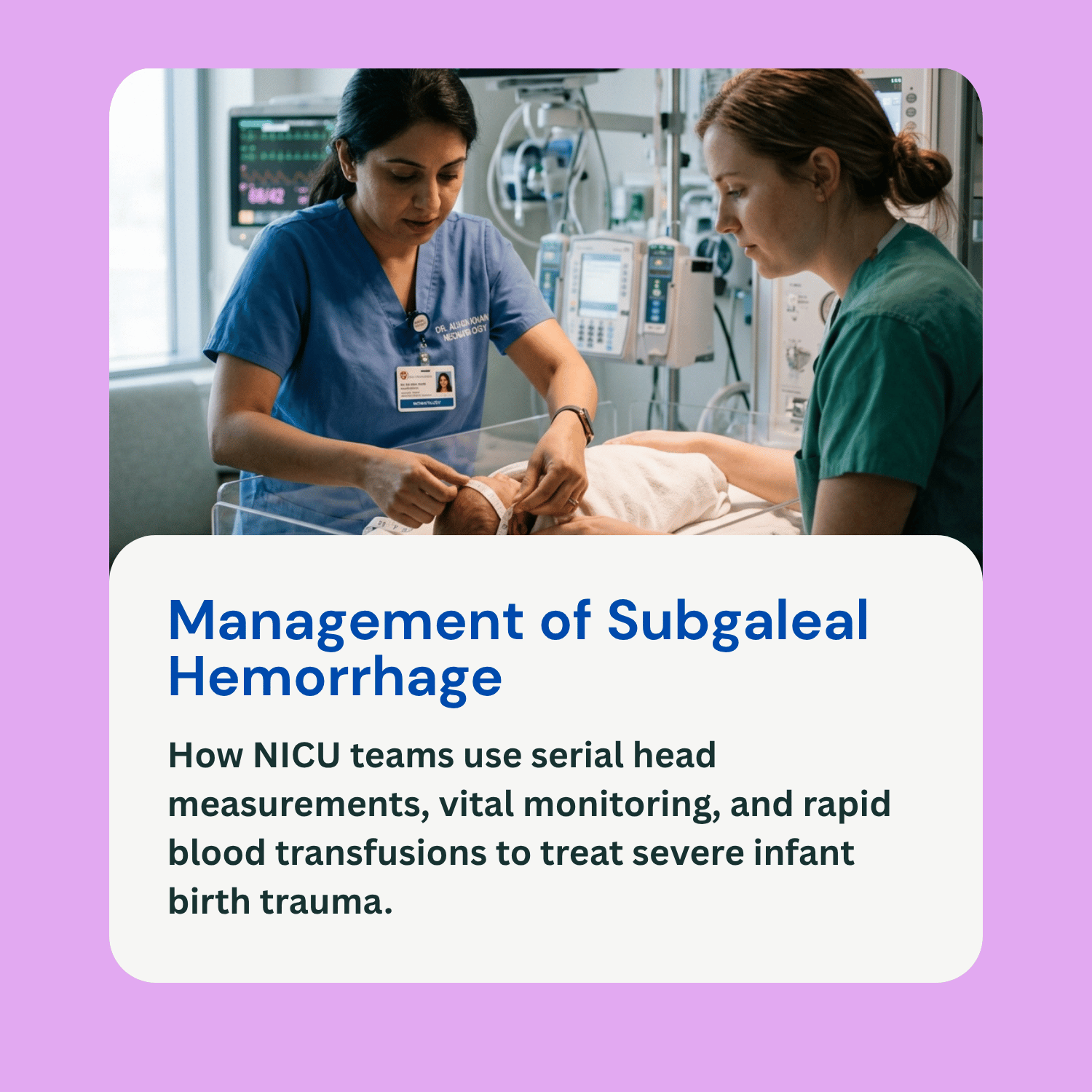
For any newborn delivered via vacuum extraction or difficult forceps application, clinical protocols recommend close monitoring—including hourly vital sign checks and serial head circumference measurements—for at least 8 hours post-delivery.
Diagnostic Evaluation: Medical teams diagnose a subgaleal hemorrhage primarily through serial physical examinations, tracking changes in head circumference, and monitoring decreasing hematocrit and hemoglobin levels. When necessary, imaging tests such as head CT scans, MRIs, or ultrasounds confirm the diagnosis and rule out structural skull fractures or intracranial brain bleeding.
Emergency Volume Resuscitation: Treatment is an emergency medical priority focused on stabilizing circulation and stopping blood loss. The baby is admitted to the Neonatal Intensive Care Unit (NICU) for immediate volume resuscitation using packed red blood cells, fresh frozen plasma, and intravenous crystalloid fluids to treat hypovolemic shock.
Coagulopathy Correction and Surgery: Massive blood loss often depletes clotting factors, leading to disseminated intravascular coagulation (DIC). Doctors administer plasma, platelets, and vitamin K to correct bleeding disorders. In rare situations where active bleeding does not stop despite aggressive medical management, emergency surgical interventions may be considered.
Potential Complications and Long-Term Prognosis
Subgaleal hemorrhage carries a high mortality rate—estimated between 12% and 25%—if not recognized and treated rapidly. However, with early detection and immediate blood transfusions, many infants recover fully. Potential short- and long-term complications include:
Severe Hyperbilirubinemia and Kernicterus: As the massive trapped blood collection breaks down over subsequent weeks, high concentrations of bilirubin enter the bloodstream. Without aggressive phototherapy or exchange transfusions, extreme jaundice can cause kernicterus, a rare form of permanent brain damage.
Hypoxic-Ischemic Encephalopathy (HIE) and Brain Injury: Prolonged severe hypotension and low blood volume deprive the brain of vital oxygen and blood supply (ischemia). This secondary hypoxic injury can lead to long-term neurological conditions such as cerebral palsy, developmental delays, learning disabilities, and seizure disorders.
Frequently Asked Questions (FAQ)
How is a subgaleal hemorrhage different from a cephalohematoma?
A cephalohematoma is a blood collection underneath the skull’s periosteum, meaning the bleed is strictly bounded by individual skull bones and cannot cross suture lines. A subgaleal hemorrhage occurs above the periosteum in a much larger tissue layer, allowing blood to freely cross suture lines, spread across the entire head, and cause life-threatening systemic blood loss.
Can a subgaleal hemorrhage be drained with a needle?
No. Doctors strongly avoid needle aspiration or surgical drainage of an uncomplicated subgaleal hemorrhage because breaking the skin creates a major pathway for bacterial entry, putting the infant at high risk for life-threatening scalp infections, abscesses, or osteomyelitis.
How quickly do symptoms of a subgaleal hemorrhage appear?
While some infants display pallor or swelling within minutes of birth, bleeding into the subgaleal space is often gradual. Symptoms may develop progressively over the first 4 to 24 hours of life, which is why close NICU observation following difficult deliveries is so important.
Can medical malpractice cause a subgaleal hemorrhage?
While subgaleal bleeding can rarely occur spontaneously, the majority of cases result from improper use of vacuum extractors or forceps. If a medical provider applies excessive force, makes too many extraction attempts, or fails to monitor the newborn for head swelling following an assisted birth, it may constitute medical negligence.