
What Causes Umbilical Cord Compression During Labor?
Throughout pregnancy and labor, the umbilical cord serves as a newborn’s life support line. Connecting the baby directly to the placenta, this vital structure transports oxygenated blood, nutrient-rich plasma, and fluids necessary for fetal survival. Under normal conditions, the soft, gelatinous tissue surrounding the blood vessels—known as Wharton’s jelly—protects the cord from flattening during routine movements and uterine contractions.
However, during labor and delivery, mechanical forces inside the uterus can squeeze or pinch the umbilical cord. This common complication is known as umbilical cord compression. When the cord is compressed, blood flow and oxygen delivery to the fetus drop immediately. While brief episodes of light compression during contractions are relatively common and often harmless, severe or prolonged compression halts the oxygen supply, leading to critical birth complications. This guide breaks down the primary causes of cord compression, how medical teams monitor for it, necessary emergency interventions, and potential birth injuries.
Primary Causes of Umbilical Cord Compression
Umbilical cord compression occurs when physical pressure inside the womb flattens the cord against the baby’s body or the uterine wall. One frequent cause is a nuchal cord, which occurs when the cord wraps around the infant’s neck one or more times. As the baby descends through the birth canal during active labor, the cord pulls tight, restricting its own blood flow. Similarly, a true knot in the cord—formed during early pregnancy as the fetus moves around—can tighten significantly during labor contractions, cutting off circulation like a kinked garden hose.
Another critical trigger is a prolapsed umbilical cord, an acute obstetric emergency where the cord slips through the cervix into the birth canal ahead of the baby. As the infant moves down during delivery, their head or body crushes the cord directly against the mother’s pelvic bones.
Fluid levels and position also play major roles. Oligohydramnios, or low amniotic fluid, deprives the cord of its natural fluid cushion, making it far more vulnerable to getting pinched between the baby and the uterine wall during every contraction. Furthermore, abnormal fetal presentations, such as breech or transverse positions, increase the risk of the cord becoming tangled or trapped under pressure.
Diagnosing Compression: Electronic Fetal Monitoring
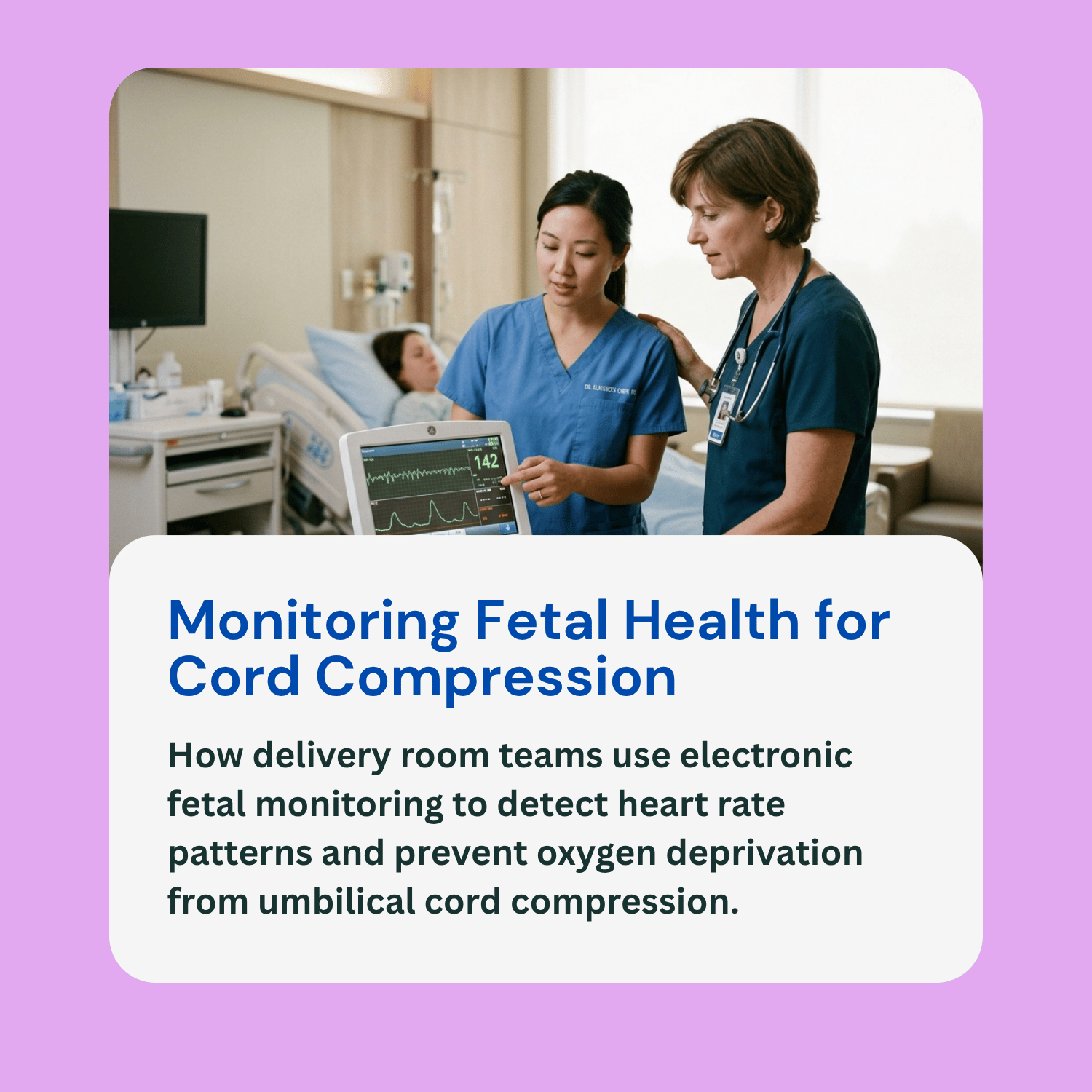
Medical teams diagnose umbilical cord compression in real time by carefully analyzing electronic fetal monitoring (EFM) printouts. Because clinicians cannot visually see the cord inside the uterus during labor, changes in the baby’s heart rate pattern serve as the primary diagnostic signal.
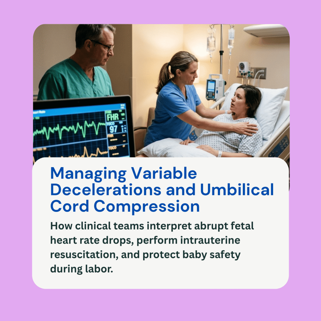
The hallmark indicator of cord compression on a monitoring strip is a variable deceleration. Variable decelerations appear as sudden, sharp drops in the fetal heart rate that resemble an abrupt “V” or “U” shape on the monitor screen. Unlike early decelerations (which occur naturally as the head compresses during contractions) or late decelerations (which indicate placental issues), variable decelerations drop rapidly and vary in duration, depth, and timing relative to contractions.
When variable decelerations are mild and brief, the baby typically recovers quickly once the contraction ends. However, if the heart rate drops deeply and stays low for extended periods—known as prolonged or severe variable decelerations—it signals that the baby is experiencing profound oxygen loss and is exhausting their physiological reserves.
Emergency Interventions in the Delivery Room
When electronic fetal monitors show signs of severe or persistent cord compression, the labor team must act immediately to restore blood flow and oxygen to the baby. Initial bedside steps focus on relieving pressure off the cord through physical positioning. Nurses will instruct the mother to shift positions—often moving her onto her left side, right side, or into a knee-chest position—to shift the fetus away from the cord.
If position changes fail to resolve the heart rate drops, doctors may perform an amnioinfusion. During this procedure, clinicians instil a warm saline solution through a small catheter directly into the uterus. This replaces missing amniotic fluid, recreating a liquid safety cushion around the cord so contractions no longer flatten it.
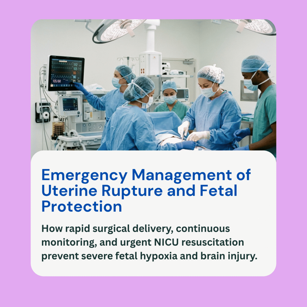
The medical team will also administer supplemental oxygen to the mother, give IV fluids, and pause labor-inducing medications like Pitocin to slow down contractions and give the baby time to rest. If these conservative measures do not rapidly restore a reassuring heart rate, or if the situation involves a prolapsed cord, the definitive intervention is an immediate emergency C-section to deliver the baby before brain injury occurs.
Potential Complications and Long-Term Birth Injuries
When umbilical cord compression is recognized and managed promptly, most babies are delivered safely with no lasting health problems. However, if medical staff miss critical variable decelerations, fail to reposition the mother, or delay an emergency delivery, prolonged oxygen debt can lead to severe, lifelong birth injuries.
Unaddressed cord compression is a major cause of birth asphyxia and Hypoxic-Ischemic Encephalopathy (HIE), a type of brain tissue damage caused by severe oxygen deprivation around birth. If HIE damages the motor processing centers of the brain, the child may develop cerebral palsy (CP), resulting in permanent impairments to muscle tone, posture, and physical mobility. Severe oxygen loss can also cause intellectual disabilities, speech delays, vision and hearing deficits, or chronic childhood seizure disorders.
Frequently Asked Questions (FAQ)
Can umbilical cord compression be detected before labor begins?
Sometimes. Standard prenatal ultrasound imaging can identify low amniotic fluid levels (oligohydramnios) or show a nuchal cord wrapped around the baby’s neck. However, because a baby frequently shifts position, cord compression is most accurately diagnosed during active labor through continuous electronic fetal monitoring.
Is a nuchal cord always dangerous?
No. A nuchal cord (cord around the neck) is actually present in up to one-third of all healthy births. In most cases, the cord remains loose enough that blood flows freely, or the doctor simply slips the loop over the baby’s head during delivery. It only becomes dangerous if it is wrapped tightly enough to restrict blood flow during contractions.
How quickly must a baby be delivered during a cord prolapse?
A prolapsed cord is an immediate medical emergency. Because the baby’s body directly crushes the cord in the birth canal, blood flow is almost completely blocked. Doctors typically perform an emergency C-section within minutes, with a nurse or doctor manually holding the baby’s head off the cord until the surgeon delivers the baby.
Can umbilical cord compression cause birth injuries due to medical negligence?
Yes. While the physical squeezing of the cord is a natural complication, failing to respond to it can constitute medical malpractice. If labor nurses miss persistent variable decelerations, fail to alert the attending physician, or delay ordering an emergency C-section when a baby is in clear distress, the resulting oxygen deprivation and brain damage may be considered preventable medical errors.