
What Is Placental Insufficiency?
Throughout a healthy pregnancy, the placenta serves as an essential, temporary organ that connects the mother’s circulatory system to her growing baby. Attached to the uterine wall, the placenta acts as a lifeline—filtering oxygen, nutrients, and antibodies from the mother’s blood and transferring them through the umbilical cord to the fetus, while simultaneously carrying away waste products.
However, when the placenta fails to develop properly or suffers damage during pregnancy, its ability to transport oxygen and nutrients becomes compromised. This serious vascular condition is known as placental insufficiency (or placental dysfunction). When the placenta cannot deliver a steady, sufficient supply of blood to the fetus, the baby’s growth and overall health are put at immediate risk. This guide breaks down the primary causes, risk factors, diagnostic methods, monitoring strategies, and long-term effects on a developing child.
Causes and Risk Factors of Placental Dysfunction
Placental insufficiency occurs when uterine blood vessels do not properly enlarge or when the placenta suffers structural damage. While it can develop unexpectedly in any pregnancy, several underlying health conditions and external factors significantly increase the overall risk.
Hypertensive disorders represent the leading cause of placental dysfunction, as chronic high blood pressure, gestational hypertension, and preeclampsia constrict the maternal blood vessels feeding the placenta, severely restricting oxygenated blood flow. Maternal vascular and metabolic conditions—such as type 1 or type 2 diabetes, kidney disease, blood clotting disorders like thrombophilia, and autoimmune diseases like lupus—can also damage micro-vessels inside the tissue. Physical placental abnormalities, including placental abruption, placenta previa, or an abnormally small placenta, directly impair gas and nutrient exchange. Furthermore, lifestyle factors such as smoking, substance use, severe malnutrition, or maternal age over 35 can restrict blood flow and alter vascular development. Finally, in post-term pregnancies extending past 41 to 42 weeks, the placenta undergoes natural aging, where calcification and tissue breakdown reduce overall efficiency.
Diagnosing and Monitoring Placental Insufficiency
Placental insufficiency often develops silently without obvious physical symptoms for the mother. However, obstetricians rely on routine prenatal screenings and targeted diagnostic tools to detect early signs of placental failure.
Measuring Fundal Height: At routine prenatal visits, doctors measure the mother’s abdomen to assess fundal height. If the uterus measures significantly smaller than expected for the baby’s gestational age, it triggers further investigation for fetal growth issues.
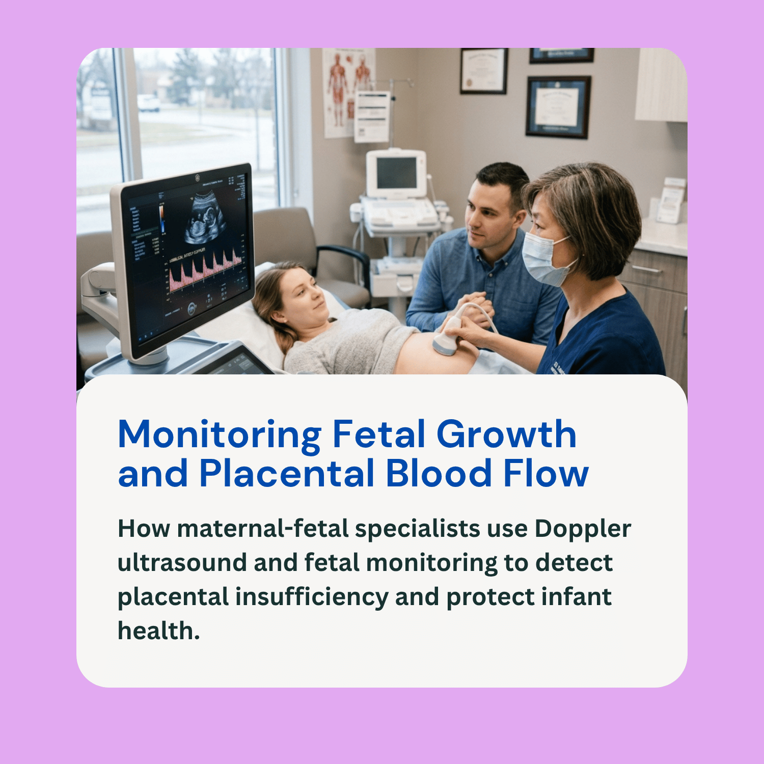
Targeted Ultrasound Imaging: Ultrasound scans allow clinicians to estimate fetal weight and assess anatomical growth. When placental insufficiency causes a baby to fall below the 10th percentile in estimated fetal weight, the condition is formally diagnosed as Intrauterine Growth Restriction (IUGR) or Small for Gestational Age (SGA).
Umbilical Artery Doppler Ultrasonography: Doppler ultrasound serves as the primary tool for measuring actual blood flow through the umbilical cord. By evaluating the resistance in the umbilical artery, doctors assess whether the placenta is allowing adequate blood flow to pass from mother to baby. High resistance, absent end-diastolic flow, or reversed end-diastolic flow indicate severe, dangerous levels of impairment.
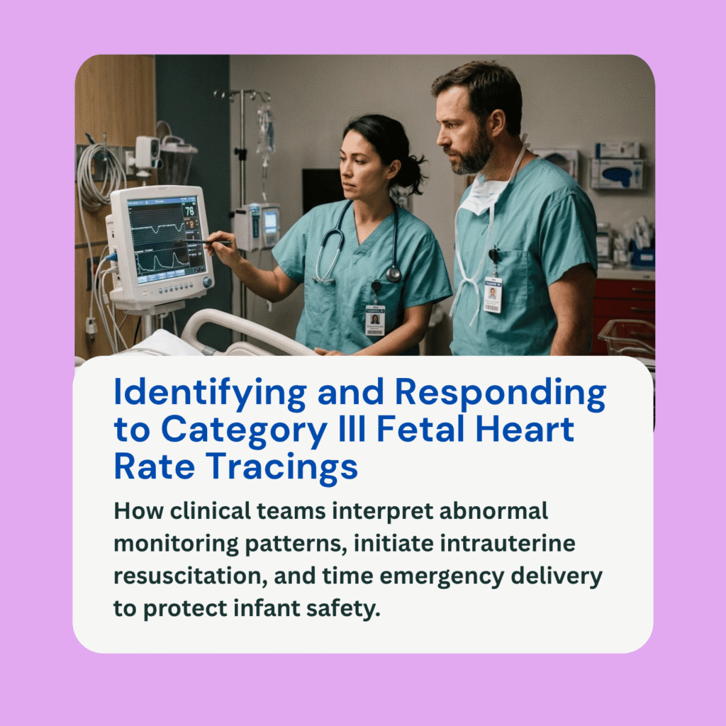
Fetal Monitoring and Biophysical Profiles (BPP): Women diagnosed with placental dysfunction undergo frequent Non-Stress Tests (NSTs) to evaluate fetal heart rate variability and response to movement. A Biophysical Profile combines an NST with an ultrasound assessment of fetal breathing, body movement, muscle tone, and amniotic fluid volume. Low amniotic fluid, known as oligohydramnios, is a common byproduct of placental insufficiency, as the baby prioritizes blood flow to the brain instead of the kidneys.
Treatment Options and Clinical Management
There is no medical cure to repair an ailing placenta once damage has occurred, so treatment focuses entirely on managing maternal risk factors and carefully timing the baby’s delivery.
Doctors work to stabilize maternal blood pressure through medications while advising modified rest or reduced physical exertion to optimize uterine blood flow. High-risk pregnancies require increased monitoring, including frequent ultrasound scans every one to two weeks alongside bi-weekly Non-Stress Tests to track fetal well-being continuously. If monitoring indicates the baby may need to be delivered prematurely, doctors administer antenatal corticosteroid injections to the mother to speed up the development of the infant’s lungs. Ultimately, the definitive treatment for severe placental insufficiency is delivering the baby. Obstetricians weigh the risks of prematurity against the risks of remaining inside an unsupportive womb, ordering an early C-section or induced delivery if Doppler studies show absent blood flow or if fetal heart rate monitors show signs of distress.
Potential Effects on the Baby
The overall impact of placental insufficiency depends on how early in pregnancy it develops and how severely blood flow is restricted.
Infants born after experiencing placental insufficiency face short-term risks such as low birth weight, hypoglycemia, hypothermia, polycythemia, and breathing difficulties due to underdeveloped lungs. Over the long term, chronic oxygen and nutrient deprivation can cause severe birth asphyxia and Hypoxic-Ischemic Encephalopathy (HIE), an acute brain injury. Affected infants face higher risks of cerebral palsy, developmental delays, learning disabilities, and chronic cardiovascular or metabolic issues later in life.
Frequently Asked Questions (FAQ)
Can a mother feel signs of placental insufficiency?
Usually, no. Placental insufficiency does not cause physical pain or obvious symptoms for the mother. The primary warning sign a mother might notice is a sudden decrease or slowdown in fetal movement as the baby conserves energy due to low oxygen levels.
Can placental insufficiency be prevented?
While structural placental problems cannot always be prevented, managing pre-existing conditions—such as keeping blood pressure controlled before and during pregnancy, avoiding smoking, and attending all routine prenatal visits—significantly lowers the risk.
What is the difference between IUGR and placental insufficiency?
Placental insufficiency is the underlying cause involving the dysfunction of the placenta, while Intrauterine Growth Restriction (IUGR) is the physical result, where the fetus fails to grow at a normal rate because of that dysfunction.
Does placental insufficiency always mean an automatic C-section?
Not always, but it significantly increases the likelihood. If the fetus shows good heart rate variability and Doppler blood flow remains stable, a closely monitored vaginal induction may be attempted. However, if the baby displays distress or if blood flow is dangerously impaired, an immediate C-section is required.